A passion for algea
Illuminated by the microscope, the green algae look like small floating jewels. In our lakes, they float as tiny plankton close to the surface and use the sunlight for photosynthesis. When algae with silicon shells die, they fall to the bottom, where their shells are stored for millennia.
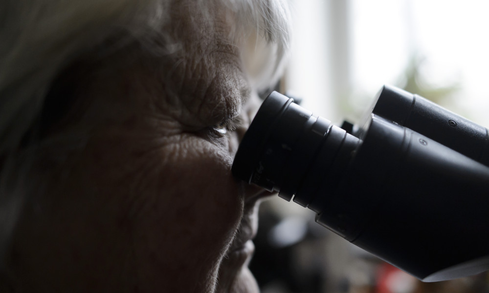
Gertrud Cronberg’s passion is studying the tiny plankton algae that are found in lakes. As a child, she developed a fascination with the life that could be discovered teaming under a microscope. Her interest led to a research career at Lund University, specialising in plankton. But why plankton algae in particular?
“They are so incredibly beautiful and there are so many different types.”
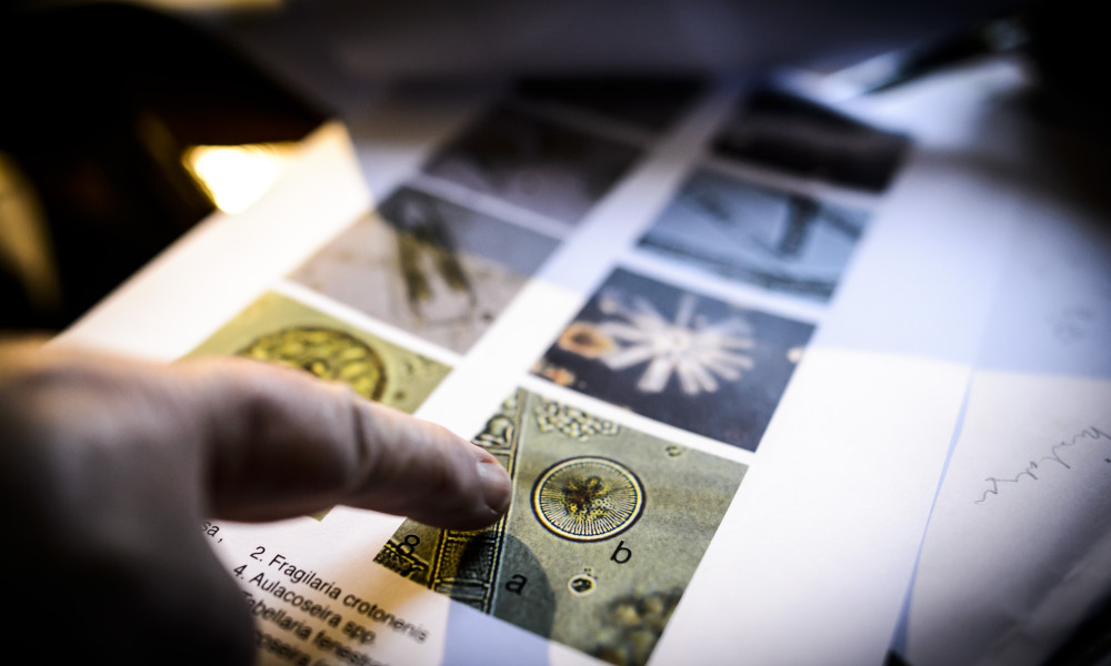
In her office at her home near Malmö, she has all the equipment she needs to view and photograph the micro-scopic algae, and she now has over 10 000 pictures in her archive. Gertrud Cronberg explains that her passion has taken her all over the world, where she has participated in many projects and collected samples from probably over 1000 lakes.
“In every water sample I recognise algae I have seen before, but sometimes I find something completely new, which is exciting!”
Text: Pia Romare
Photo: Kennet Ruona and Gertrud Cronberg
Published: 2014
Facts
-
Image 3 shows
-
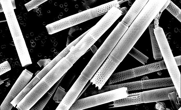
Two different species of silicon algae, also known as diatoms, of the genus Aulacoseira. The diameter of the cells on the picture is approximately 20–30 µm. This genus includes many species and is present worldwide. Its cells are covered with a silicon shell and the shell of every species has a unique appearance. The silicon shell is very resistant and is preserved in lake sediment for tens of thousands of years, storing information about the climate and environment of the area. They are present in plankton, especially during the spring and autumn in nutrient-rich lakes.
The picture was produced by Dr Gertrud Cronberg, Aquatic Ecology, Lund University using a scanning electron microscope.
-
Image 4 shows
-
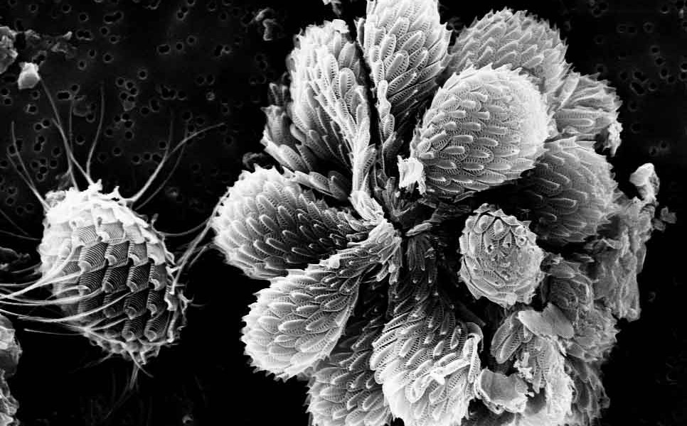
The silicon-shelled golden algae Synura petersenii (the large colony in the centre of the image) and Mallomonas Striata (on the right). The Synura colony is approximately 50 µm across and the Mallomonas colony is 25 µm long. Their cells are covered with silicon shells and the shell of every species has a unique appearance. All of the cells also have flagella with which they swim and keep themselves afloat. These golden algae are present in moderately nutrient-rich lakes.
The picture was produced by Dr Gertrud Cronberg, Aquatic Ecology, Lund University using a scanning electron microscope.
-
Image 5 shows
-
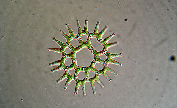
A colony of green algae Pediastrum duplex variant gracillimum, with a size around 100µm. In the star-shaped colony all green cells have their own particular place. These green algae are generally present in plankton, but also anchor to aquatic plants. In the sediment the cells of the genus Pediastrum are well preserved because there is silicon in the cell walls. Thus, Pediastrum is often used as a guide fossil when studying the historical development of a lake, for example from a nutrient-poor to a nutrient-rich environment.
The picture was produced by Dr Gertrud Cronberg, Aquatic Ecology, Lund University using an optical microscope.
-
Image 6 shows
-
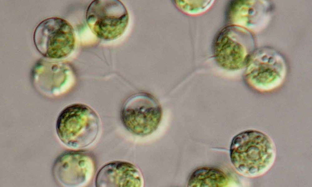
A colony of green algae Dictyosphaerium pulchellum. The colony comprises green cells that sit on thin branches in groups of four, and the size of the entire colony is around 50µm. These algae are present during the warmer seasons of the year in moderately nutrient-rich lakes and can often be found together with blue-green algae.
The picture was produced by Dr Gertrud Cronberg, Aquatic Ecology, Lund University by using an optical microscope with transmitted light illumination.


